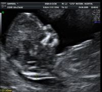
 The ultrasound scan at 11-14 weeks consists the first detailed examination of the fetus and allows the calculation of the probability of chromosomal abnormalities, of which one of the most common is Down's syndrome or mongolism. Children with Down’s syndrome always have to some degree mental retardation while often have heart problems. Any woman can give birth to a child with Down’s syndrome, but the probability increases with increasing age.
The only way to be certain that the fetus has not gotten Down’s syndrome is by an intervention (amniocentesis or chorionic villus sampling), which examines the genetic material from the amniotic fluid (amniocentesis) or the placenta (chorionic villus sampling) that is identical to the genetic material (chromosomes) of the embryo.
However, these interventions have a small percentage of miscarriage (about 1%). For this reason, the decision whether or not to proceed to such an intervention is best based on a test that calculates the probability of the fetus having Down’s syndrome. This test is the ultrasound examination at 11-14 weeks where the measurement of nuchal translucency is performed and the presence of the nasal bone (the bone in the nose of the fetus) is evaluated. These findings are combined with the measurement of two hormones in the blood of the mother (PAPP-A, b-hCG). The nuchal translucency is a liquid found in the fetal neck, underneath the skin. All embryos have this fluid, but those suffering from Down’s syndrome usually have more liquid. During the ultrasound scan performed at 11-14 weeks the growth and the major organs of the fetus are also being monitored. By measuring the nuchal translucency, nasal bone and the two hormones in the mother's blood, we can predict at least 9 in 10 children suffering from Down’ Syndrome (95% sensitivity).
The result that you will eventually get is a number that represents your personal chance of having a baby with Down’s syndrome. The result is based upon your age, your medical history (it is important to state whether you had previous pregnancies with a similar problem or have a family history), the fetal measurement and the hormones in your blood. For example, if your score indicates probability 1:2000, this means that if we had 2000 women like you, one would have a fetus with Down’s syndrome and the rest 1999 would have had normal embryos. The examination at 11-14 weeks can never assure you that the fetus has not gotten Down’s syndrome.
This method of calculating the probability of Down’s syndrome is very precise and its results have been confirmed in hundreds of thousands of women worldwide. In addition, the department of Ultrasonography and Embryo-Maternal Medicine of LETO Maternity Hospital is staffed by physicians who have the necessary certificates of competence from the Fetal Medicine Foundation (FMF, www.fetalmedicine.com) in London which developed this method. FMF verify the results of all internationally recognized centres in order to maintain the same high level.
The results of the ultrasound and biochemical testings will show low risk for Down’s syndrome (i.e. probability of less than 1:300) in most women.
If your score indicates greater likelihood than 1:300, it means that you should consult your doctor and together decide whether to proceed surgically (amniocentesis or chorionic villus sampling) to check with certainty the chromosomes of the fetus. Even in high-risk groups most embryos are in the end normal.
Thus, for example if the fetus has a probability of 1:100 of Down’s syndrome, this means that out of 100 pregnancies only one would result in a problematic fetus, while the remaining 99 would be normal.
The fetuses with increased nuchal translucency and normal chromosomes are usually normal. However, they have greater probability for congenital abnormalities of various organs especially heart disease and some rare diseases (genetic syndromes). Many of these problems can be screened with detailed ultrasounographic imaging and that is why those embryos require more specialized testing.
 With this ultrasound the gestational age is calculated with precision, which is especially important to women with unstable menstrual cycles. In multiple pregnancies, the chorionicity is estimated, i.e. whether the fetuses have separate placentas or a common placenta, information which determines the course of the pregnancy and how to monitor it. Finally, this ultrasound scan offers the possibility of a first rough estimation of fetal anatomy as to be able to detect a significant proportion of serious abnormalities at this early stage.