The 3D ultrasound (3D and 4D)
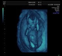 Technology now allows us to have three-dimensional imaging of the fetus. The three-dimensional image (3D) occurs from the composition of many two-dimensional images. In the more sophisticated devices the fetus can be seen three-dimensionally in real time (4D), i.e. to see his/her movements.
The usual two-dimensional ultrasonographic image gives us the most important information about the fetus. In some rare abnormalities the three-dimensional ultrasound scan can help us see certain organs better, however, the two-dimensional ultrasound basically guides us in our diagnosis.
The biggest advantage of three-dimensional ultrasonography is that it gives us the satisfaction to see ‘truer’ images of the fetus in his/her natural environment.