Colour Doppler ultrasound scan
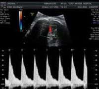 With the colour Doppler ultrasound the blood flow in the uterine arteries is monitored. If the resistance in the uterine arteries is high the likelihood of the mother developing pre-eclampsia or the fetus having little weight increases. Approximately one in four women with increased resistance will have such problems, while in the rest of the female population the pregnancy will develop normally. If during the B level ultrasound scan increased resistance is found close monitoring of the pregnant woman’s blood pressure and monthly ultrasound scans of the fetus are recommended.
Finally in the B-level ultrasound the length of the cervix is being measured. The cervix is the lower part of the uterus that opens up (expands) in childbirth. The measurement is done reliably by trans-vaginal ultrasound. If the cervix is significantly smaller (less than 15mm), the risk of premature birth increases. Recent studies have shown that by providing better treatment to women with short cervixes, the incidence of extremely premature birth is reduced by 40%.
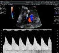 The growth ultrasound scan is performed starting at 24 weeks until the end of pregnancy, depending on the physician’s advice. Fetal growth, weight, quantity of amniotic fluid, the location of the placenta are being monitored. The Doppler examination is the control of the blood flow of the embryo’s vessels and placenta (usually the flow in the umbilical artery and the middle cerebral artery is examined), which gives us information on the functioning of the placenta and the status of the fetus. This information is particularly important for small weight fetuses and helps us decide whether an underweight (small for gestational weeks) fetus receives enough oxygen and nutrients from the placenta. In cases where the flow in these vessels is not normal more detailed and frequent monitoring is required.